


 02 9541 3500
Dental Applications of Scanning Electron Microscopes
What are scanning electron microscopes (SEMs)?
Where traditional microscopes use light waves, scanning electron microscopes (SEMs) use a beam of electrons to scan a sample’s surface and produce images of high magnification with a great deal of topographical information. Micrograms produced by SEMs are critical in research, development and ongoing improvements for dental and orthodontic practice.
The Phenom desktop SEM
The Phenom ProX SEM is a tabletop scanning microscope able to provide magnification of up to 150 000x . This powerful capability is combined with the ease of use of a traditional microscope and Phenom SEMs fit comfortably into most laboratories and dental surgeries. They are most suitable for universities and research, design and manufacturing facilities, where high magnification imaging and ease of use are foundational requirements.
Research and development applications
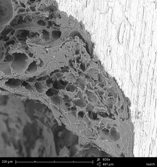
Studying dental wear with SEMs
In 2014 the Department of Surgical and Morphological Sciences at Insubria University in Varese, Italy conducted a study on four different types of dental wear:
- Erosion
- Attrition
- Abrasion
- Abfraction
The three scientists that conducted the study, Luca Levrini, Giulia Di Benedetto and Mario Raspanti, all from the University’s Oro Cranio Facial Disease and Medicine Research Centre, used a scanning electron microscope to ‘clarify the different clinical and diagnostic presentations of dental wear and their possible significance’.
They found that each lesion type had specific morphological and etiological factors and that identifying these factors allowed for the identification of complex nondental disorders, such as acid reflux. This experiment highlights the benefits of using an SEM in dental research.
Wire bending and salivary pH on surface areas
Scanning Electron Microscopes were a key measuring tool in an experiment conducted by Progress in Orthodontics into the effect of saliva pH on dental wiring. In this study, an SEM was used to observe:
- A control group
- Bent wiring exposed to artificial saliva
- Straight wiring exposed to artificial saliva
The SEM’s high magnification allowed observation of the differences between the control group and the wiring exposed to saliva pH. In particular, the SEM allowed the experimenters to differentiate between striations caused in the manufacturing process of the writing and surface irregularities.
The experiment was a joint study between The Orthodontic Department of the College of Medicine and Dentistry at James Cook University, the School of Dentistry at the University Medical Centre Groningen and the School and Hospital of Stomatology at Wenzhou Medical University. They found that bending has ‘a significant influence on surface roughness and mechanical properties of rectangular SS archwires’ and that ‘pH has a synergistic effect on the change of mechanical properties of stainless steel (SS) wires along with wire bending.’
SEM in depth profiling PET analysis
An article in American Laboratory (via ResearchGate) used scanning electron microscopes to examine the changes in fibre morphology upon SVI treatment at a variety of temperatures.
The purpose of this experiment was to examine ways bulk polymers could be functionalised without modifying their potential for electronic or optical purposes. AFM-IR, which combines atomic force microscopy and infrared spectroscopy, allowed for measurements of the depth of precursors into fibres, with the aim of shortening development and processing times.
Without the use of an SEM, such depth of analysis of the polyethylene terephthalate fibres would not have been possible.
Evaluating the bond strength of dental base brackets using SEMs
A research project conducted by the University of Messina evaluated the bond failure of metal brackets bonded to enamel.
The paper, titled ‘Evaluation of Bond Strength and Detachment Interface Distribution of Different Bracket Base Designs’ and published in Acta Medica Mediterranea, showed through the use of scanning electron microscopes that brackets with greater mesh spacing had the best bond results, with double layer mesh patterns in 80/150 gauge double mesh having the best bond patterns. In this research, SEM usage aided the researchers in identifying the types of mesh that were effective for enamel bonding.
Using SEMs to chart the effects of solvent evaporation
In dental and orthodontic treatments, the effects of solvent evaporation on water sorption and solubility can have significant effects on the restorative techniques of dentin bonding materials.
An experiment conducted by the School of Dentistry at the University of Brasilia used SEM micrographs to examine nanoleakage patterns prior to, and after evaporation. Conducted over a series of days, the micrographs allowed researchers to compare and contrast different solvents under different conditions. This kind of high magnification analysis would not be possible without an SEM.
SEMs in dental and orthodontics
Scanning electron microscopes are an indispensable tool in dental and orthodontic research and development. The capacity for high magnification analysis is particularly critical to improve health, aesthetics, cost and function of materials and techniques.
The current magnification for the Phenom Pharos SEM can reach up to 1,000,000 x. !!
Phenom provides an all in one imaging and analysis system. Elemental analysis is built into the desktop system to give point and click element identification. The Phenom desktop SEM is extremely fast and easy to operate taking just 30 seconds to display an SEM image after loading a sample.
Contact us for a quote.